Pelvic Region
pelvic region of the trunk is located in the posterior and inferior to the abdomen and is considered the place of transition of the trunk and extremities (1). It is composed of bones, ligaments and muscles (1). The bones that make up the pelvic region are known as pelvic girdle (1), pelvic or hip (2).
The perineum is the area of \u200b\u200bthe trunk between the thighs and buttocks, stretching from the coccyx to pubic (1). In men, this area contains the penis, scrotum and anus, while in women includes the external genitals and the anus (1).
Bones of the pelvic girdle
The pelvic girdle consists of two coxal bones that join in front by the pubic symphysis and behind by the sacroiliac joint (2). The complete ring is formed provides a solid and stable spine and pelvic organs (2), such as the bladder, the terminal portions of the ureters, genitalia and rectum (1).

Each innominate bone is composed of three separated by cartilage called the ilium, ischium and pubis (1) (2). The ilium is located in the upper region, the pubis in the inferior and anterior ischium is finally in the inferior and posterior (2). Figure 1 shows a diagram of these bones, including the femur (thigh bone) and iliac crest (upper region of the ilium). Although these three bones fuse around 23 years, usually studied separately (2). Figure 2 shows the acetabulum cavity where the femoral head articulates (2).
 Figure 2: Acetabulum
 http://www.anatomiavet.cl/2009/07/acetabulo-hueso-acetabular.html
The general structure of the male and female pelvis has some very obvious differences (1) (2), mainly due to the adjustments related to pregnancy and childbirth.
The male pelvis is hard and heavy, small pelvic ring heart shaped, the acetabulum is large and looks to the side and the pubic arch is less than 90 degrees. The female is lighter and slim, the pelvic ring is large and oval and the acetabulum is smaller and tends to look before. The pubic arch has an angle of 90 degrees (1) (2).
The bony pelvis is divided into an upper and a lower portion across the boundary line marked by the terminal shown in Figure 3. The upper part is called greater or pelvis false pelvis, while the lower pelvis is known as lower or true pelvis (2).

Figure 3: Line Terminal
http://www.esacademic.com/pictures/eswiki/83/Skeletpelvis-pubis.jpg
http://www.esacademic.com/pictures/eswiki/83/Skeletpelvis-pubis.jpg
The Femur
The femur, thigh, is the longest, heaviest and toughest of all the bones of the body (1) (2). As mentioned above, the proximal end articulates with the acetabulum and its distal end articulates with the tibia and patella or patella (2). The body or shaft of the femur is inclined medially and this feature is more prominent in women as they have wider pelvic region (2).
Figure 4 shows the main parts of the femur. At the proximal end is the head, neck, trochanter major and minor (2). These two are projections arising from the junction of the neck to the body and act as insertion site for different tendons of the thigh and gluteal region (2), among them is the intertrochanteric line, it is important to mention it is a common site where there are hip fractures. The greater trochanter can be felt and seen in front of depression on the sides of the hip (2).
At the lower end of the femur was located medial condyle and lateral condyle, which articulates with the tibia and is inserted where the ligaments of the knee (2).

Hip Joint
The hip joint, also called hip joint is formed by the head of the femur and acetabulum of the innominate (2). It consists of a joint capsule very dense and strong, and a group of ligaments that allow flexion, extension, abduction, adduction, and rotation circumduction medial and lateral thigh. Some of these components are illustrated in Figure 5.

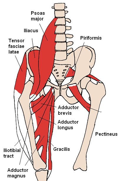
Pelvic Muscles
Most of the muscles that move the femur originating from pelvic girdle and insert on the femur. At the level of the hip joint are the following muscles and their functions (2):
- The iliopsoas flexes the thigh and assists in lateral rotation and flexion of the trunk.
- The iliacus and gluteus maximus extends and laterally rotates the thigh.
- The gluteus medius and minimus abduct the thigh and rotate medially.
- The tensor fascia lata is located on the lateral surface and its role is to flex and abduct the thigh.
- The piriformis, obturator internal and external, upper and lower twin quadratus femoris laterally rotate the thigh.
- The adductor longus, short and most argue, flex and medially rotate the thigh.
- pectineus flexes and adducts the thigh.

Figure 6: Muscles of the Pelvis, anterior
http://seeadamtrain.files.wordpress.com/2010/02/anterior_hip_muscles_21.png
http://seeadamtrain.files.wordpress.com/2010/02/anterior_hip_muscles_21.png
Nerves Pelvic Region
The sacral plexus is a collection of axons that arise from the roots previous spinal nerves L4-L5 and S1-S4 (2). Are arranged along the anterior sacrum and innervate buttocks, perineum and lower limbs. The sciatic nerve is considered the longest in the body, has its origin in this plexus (2).
Some of the major nerves of the plexus and their respective distribution shown in Figure 7 and discussed below (2):
- The Superior Gluteal supplies the gluteus minimus muscle, medium and tensor fascia lata .
- The inferior gluteal, piriformis, quadratus femoris, and Lower Twin Internal Shutter innervate the muscles of the same name.
- Internal Shutter supplies the internal obturator and superior twin
- The Sciatic, consisting of the common tibial and common peroneal nerves innervating various muscles of the leg.
- the pudenda that supplies the muscles of the perineum, skin of the penis and scrotum of the man, the clitoris, labia majora, labia minora and vagina in women.
coccygeal plexus is a small network of nerve fibers formed by the anterior rami of S4 and S5 and coccygeal nerves (1) and although born in the pelvic region, has no direct relationship with hip muscles.

Irrigation Region
Pelvic Arteries enter the pelvis from the common iliac artery to turn is a branch of the descending aorta (1). The main ones are:
- internal iliac arteries, considering the arteries of the pelvis, emit branches to the buttocks, medial thigh and perineum (1).
- median sacral artery, which supplies the last lumbar vertebrae, sacrum and coccyx (1).
- superior rectal artery, which supplies the upper portion the rectum and joins the lower and middle rectal arteries, who are branches of the internal iliac (1).
blood coming from the pelvic viscera returns to the heart via the inferior vena cava (2). The main one is the internal iliac vein, which drains not only the internal organs of the pelvis but also the thighs, buttocks and genitalia (2).

Figure
8: Arteries and Veins of the Pelvis
http://en.academic.ru/pictures/enwiki/73/Iliac_veins.gif
http://en.academic.ru/pictures/enwiki/73/Iliac_veins.gif
References
(1) Moore, Keith . Dalley, Arthur. 2003. Clinical Oriented Anatomy. Edition 4. Editorial Médica Panamericana. Argentina.
(2) Tortora, Gerald. Derrickson, Bryan. 2006. Principles of Anatomy and Physiology. 11 th. Edition. Editorial Médica Panamericana. Mexico
0 comments:
Post a Comment